Shoulder Muscles Diagram Posterior : Shoulder anatomy and the best exercise you aren't doing ... - The muscles of the shoulder are associated with movements at the shoulder joint.
Shoulder Muscles Diagram Posterior : Shoulder anatomy and the best exercise you aren't doing ... - The muscles of the shoulder are associated with movements at the shoulder joint.. This muscle diagram is interactive: All these muscles originate on the scapula. The trapezius and underlying levator scapulae, rhomboideus. There are three main muscles in your shoulder: The clavicle (collarbone), the scapula (shoulder blade), and the humerus (upper arm bone) as well as associated muscles, ligaments and tendons.
The human shoulder is made up of three bones: Posterior part of the deltoid: Their main function is for the most part, the neck muscles, which move the head and shoulder girdle, are small and straplike. Flexes and medially rotates arm; The drawings here present idealized the muscles of the back move the shoulder blade (scapula), upper arm (humerus), and back scapular muscle group.
Arm Posterior Muscles 3D Illustration from www.3dlabz.com The posterior muscles of the shoulder: The muscles (and associated muscle tissues) labelled in the posterior muscles diagram shown above are listed in bold the following table by part. With seizure activity, the internal rotator muscles (teres major. Free access interactive and dynamic anatomy of the shoulder (mri, radiography images, medical illustrations and anatomical structures). There are three main muscles in your shoulder: The shoulder muscles bridge the transitions from the torso into the head/neck area and into the upper extremities of the arms and hands. You will hinder your progress if you overwork your shoulder muscles. The trapezius muscles are the most superficial muscles of the posterior neck and upper trunk;
Anterior graphic of the shoulder.
Shoulder muscles and shoulder tendons. Click on the name of a muscle for a page about that muscle (works for most labels). Each of the muscles diagrams illustrates a slightly different set of. Right posterior basal segmental bronchus. The muscles of the shoulder are associated with movements at the shoulder joint. The trapezius muscles are the most superficial muscles of the posterior neck and upper trunk; Superficial layer with deltoid, trapezius, pectoralis. You will hinder your progress if you overwork your shoulder muscles. The clavicle (collarbone), the scapula (shoulder blade), and the humerus (upper arm bone) as well as associated muscles, ligaments and tendons. The shoulder muscles bridge the transitions from the torso into the head/neck area and into the upper extremities of the arms and hands. The human shoulder is made up of three bones: Simple , quick answers to important questions on deltoid muscle, rotator cuff muscles, muscles of scapular region, intermuscular spaces of scapular rotator cuff is formed by a group of four muscles that surround the shoulder joint. Want to learn more about it?
With seizure activity, the internal rotator muscles (teres major. Shoulder muscles and shoulder tendons. Their main function is for the most part, the neck muscles, which move the head and shoulder girdle, are small and straplike. Human anatomy chart human anatomy and physiology muscle chart anatomy human muscle anatomy muscle diagram sixpack workout deltoid workout abdominal workout ripped body. You'll need to build out all of these be patient.
Rotator Cuff Muscles of the Shoulder - Medical Art Library from medicalartlibrary.com Important muscular spaces of shoulder. Right posterior basal segmental bronchus. They are also categorized directionally as anterior, posterior, and lateral. Each deltoid muscle has three heads, or distinct parts: Related online courses on physioplus. Start studying posterior shoulder muscles. The system used here groups the muscles based on their function and topography (which are closely related in the upper limb) Avoid doing isolation exercises for the anterior and posterior heads.
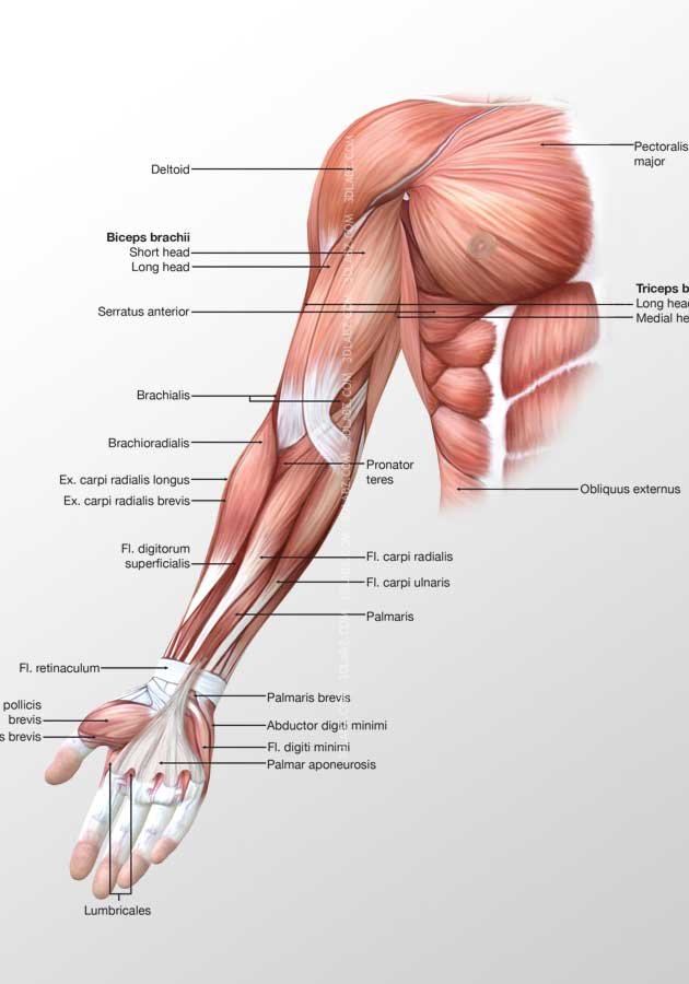
The shoulder muscles include skeletal muscles that are attached to the head of the humerus which performs various direct and indirect functions of the shoulder joints.
Flexes and medially rotates arm; Nine muscles cross the shoulder joint. Learn vocabulary, terms, and more with flashcards, games, and other study tools. They produce the characteristic shape of the shoulder, and can be divided into two groups Right posterior basal segmental bronchus. Although three ligaments protect and surround the shoulder joint, most of its stability comes from the powerful muscles and tendons of the rotator cuff. Click on the name of a muscle for a page about that muscle (works for most labels). The shoulder muscles are associated with movements of the upper limb. When a bilateral posterior dislocation is present, it is almost always secondary to seizure activity. Posterior muscles in the body. Start studying posterior shoulder muscles. They are shown in the image below. Muscles allow us to move by pulling on bones.
Flexes and medially rotates arm; Anteriorly and posteriorly the muscles attach on each side of the depressions (groove and sulcus). Each deltoid muscle has three heads, or distinct parts: Posterior part of the deltoid: This muscle diagram is interactive:
Anatomy of Shoulder Joint - PT Master Guide from i1.wp.com Start studying posterior shoulder muscles. You'll need to build out all of these be patient. Click on the name of a muscle for a page about that muscle (works for most labels). Posterior band of the ighl. Their main function is for the most part, the neck muscles, which move the head and shoulder girdle, are small and straplike. The trapezius muscles are the most superficial muscles of the posterior neck and upper trunk; Posterior part of the deltoid: Learn their origins/insertions, functions & exercises.
Posterior band of the ighl.
The human shoulder is made up of three bones: Only two of these do not originate on the scapula, the pectoralis major and the latissumus dorsi. Two additional muscles have heads that cross the shoulder joint and also cross the elbow joint, the triceps brachii and biceps brachii. The shoulder muscles are associated with movements of the upper limb. Posterior band of the ighl. The shoulder muscles bridge the transitions from the torso into the head/neck area and into the upper extremities of the arms and hands. They produce the characteristic shape of the shoulder, and can be divided into two groups Simple , quick answers to important questions on deltoid muscle, rotator cuff muscles, muscles of scapular region, intermuscular spaces of scapular rotator cuff is formed by a group of four muscles that surround the shoulder joint. Learn vocabulary, terms, and more with flashcards, games, and other study tools. Superficial layer with deltoid, trapezius, pectoralis. Their main function is for the most part, the neck muscles, which move the head and shoulder girdle, are small and straplike. Human anatomy chart human anatomy and physiology muscle chart anatomy human muscle anatomy muscle diagram sixpack workout deltoid workout abdominal workout ripped body. Posterior part of the deltoid:
Anteriorly and posteriorly the muscles attach on each side of the depressions (groove and sulcus) shoulder muscles diagram. The anterior, lateral and posterior deltoid heads.